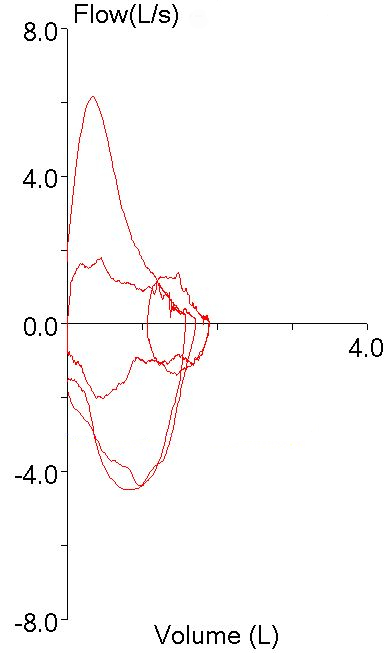
A couple of days ago I was reviewing (triaging, actually) the spirometry portion of a full panel of PFTs performed with pretty terrible test quality and was trying to decide if the technician responsible for performing the tests had made the right selections from the patient’s test results. I noticed that the FEV1 that had been selected was actually the lowest FEV1 from the all the spirometry efforts the patient made, and was trying to decide whether this was really the correct choice. We use peak flow to help determine which FEV1 to select and that particular spirometry effort appeared to have the highest and sharpest peak flow by a large margin:
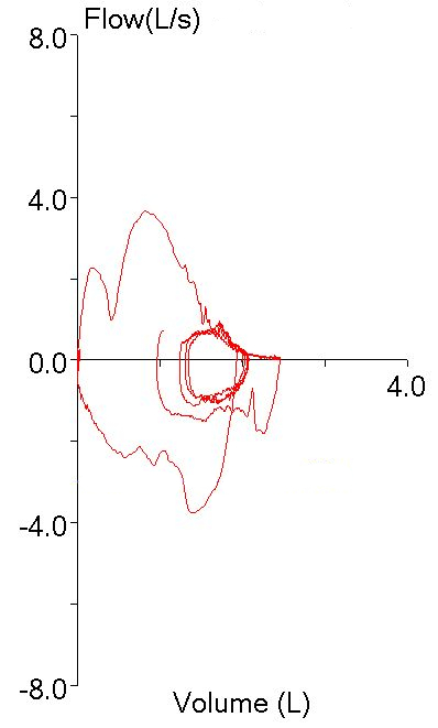
particularly when compared to the other spirometry efforts:
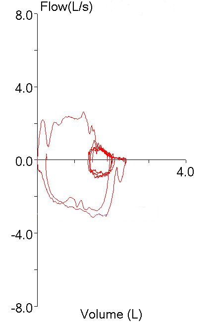
But this was hard to reconcile given how low the FEV1 was relative to the others:
| Test #1 | Test #2 | Test #3 | ||||
| Observed: | %Predicted: | Observed: | %Predicted: | Observed: | %Predicted: | |
| FVC (L): | 1.71 | 41% | 2.46 | 59% | 2.39 | 58% |
| FEV1 (L): | 1.24 | 39% | 1.81 | 57% | 1.77 | 55% |
| FEV1/FVC: | 73 | 95% | 74 | 96% | 74 | 97% |
All of the other efforts though, had significant quality issues that might cause the FEV1 to be overestimated. Effort #2 had a large amount of back-extrapolation (0.32 L, 13% of FVC). Effort #3 also had a lot of back-extrapolation (0.28 L, 12% of FVC) and it also had a blunted peak flow that was substantially lower than the other efforts. Or did it?
I was looking across all the numerical peak flow results and suddenly noticed that the first effort, the one that appeared to have the highest peak flow, actually had the very lowest.
| Test #1 | Test #2 | Test #3 | ||||
| Observed: | %Predicted: | Observed: | %Predicted: | Observed: | %Predicted: | |
| PEF (L/sec): | 1.79 | 21% | 3.66 | 0.44 | 2.64 | 0.32 |
So what was going on here?
As I said, the patient’s test quality was pretty poor and when I looked at the volume-time curve I saw what the problem was.
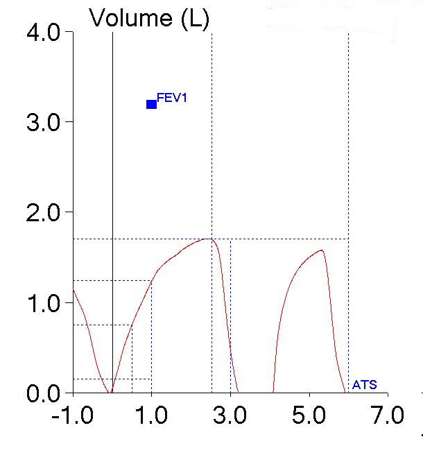
The patient had actually made two back-to-back efforts. The numerical values (FVC, FEV1 and PEF) were reported from the first effort. The second effort however, was what produced the flow-volume loop with the highest peak flow. The smaller loop that was inside the maximal loop (which I had taken to be one of the tidal loops and ignored) was actually the first effort.
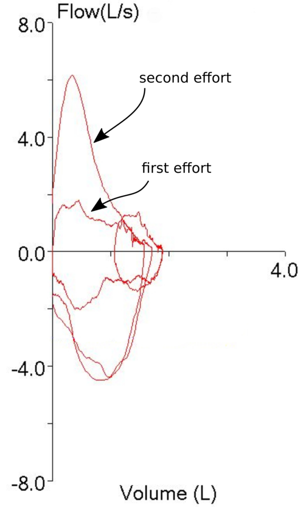
So, both the technician and I had been fooled by looking at the first effort’s flow-volume loop and taking it at face value. It was obvious that the first spirometry effort had the highest peak flow so the FEV1 from that effort had to be the best and most accurate one, didn’t it? Well actually, no, it didn’t, particularly since we weren’t looking at the right flow-volume loop.
To some extent I blame our lab’s software for this problem. The traces that make up the flow-volume loop are presented in one color with no differentiation between the pre-test tidal loops, the test effort itself, and whatever comes after the test effort. Oftentimes towards the end of an exhalation, the tracings from the tidal loops and the actual spirometry effort frequently overlap and there have been many times when I have wished that the tidal loops were in a different color in order to make it clear which tracing was which (particularly when I’m trying to determine whether the spirometry effort ended at a volume lower than where it started). This particular problem makes it clear that the tracing color should also be different once the end of a spirometry effort has occurred. Maybe if it looked something like this:

then it would be clear which part of the flow-volume loop was which.
I ended up selecting effort #2 to be reported, partly because it had the largest FVC (and longest expiratory time), partly because it had the best PEF and partly because even if the FEV1 was overestimated because of of back-extrapolation, it was still more believable than the FEV1’s from all the other efforts. I also think this was a better choice since if the FEV1 from the first effort had been reported it would have looked like the patient had airway obstruction on top of his restriction (the TLC was moderately reduced) and I just don’t think that was correct.
The fact that I was trying to salvage something out of this patient’s tests results re-raises the question of when results should be reported and when they shouldn’t. You could say that if none of the tests meet the ATS/ERS standards then none of them should be reported and that argument could be made for this patient’s spirometry efforts since none of the spirometry efforts met all of the ATS/ERS standards. The problem with taking this approach is that the ATS/ERS spirometry standards have loopholes. In particular you can take the FEV1 from one effort and the FVC from another. If this was done then the FEV1 from the first effort would have been reported since it was the only one that had essentially no back-extrapolation (0.05 L) and the ATS/ERS standards do not use PEF in any way when selecting FEV1. The effort the FVC would have been taken from was longer than 6 seconds and also met the end-of-test criteria. For these reasons it could be said that the results derived in this way would have met the ATS/ERS standard even though it’s also apparent that the test quality wasn’t particularly good and that the results probably wouldn’t reflect the patient’s status.
There’s also the point that even when test results don’t meet all of the ATS/ERS criteria, they can still answer some questions. For example if a spirometry effort was only 1-1/2 seconds long but the FEV1 was normal, then that makes it reasonably unlikely that there is any airway obstruction even if the FVC is significantly underestimated. I’d say that’s a reason to still report the results. Ditto if the FVC was above the LLN but the FEV1 was mis-estimated due to an expiratory pause or back-extrapolation.
For this patient test quality and reproducibility was poor, but there were still bits and pieces that were able to present a somewhat coherent picture and for this reason (after some editing) I put the report in my out basket.